A line through the brain
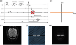
16 September 2021Functional magnetic resonance imaging (fMRI) is a widely used tool in neuroscience to detect neurally evoked responses, e.g. the blood oxygenation level-dependent (BOLD) signal. Typically, BOLD fMRI has millimeter spatial resolution and temporal resolution of one to few seconds. To study the sub-millimeter structures and activity of the cortical gray matter, the field needs an fMRI method with high spatial and temporal resolution. Line-scanning fMRI achieves very high spatial resolution and high sampling rate, at the cost of a sacrifice in volume coverage, akin to a virtual electrode sampling blood oxygen levels.
In a paper by Luisa Raimondo and colleagues, a human line-scanning implementation on a 7T MRI system is presented. First, the quality of the saturation pulses that suppress MR signal outside the line was confirmed. Second, the best coil combination for reconstruction was established. Finally, they used the line-scanning method in the occipital lobe during a visual stimulation task, showing BOLD responses along cortical depth, every 250 µm with a 200 ms repetition time (TR).
This demonstrates the feasibility of line-scanning in humans. Further research will apply line-scanning fMRI for applications in cognitive and clinical neuroscience.
Link to full paper 10.1177/0271678X211037266
Raimondo L, Knapen T, Oliveira IAF, Yu X, Dumoulin SO, van der Zwaag W, Siero JCW. A line through the brain: implementation of human line-scanning at 7T for ultra-high spatiotemporal resolution fMRI. J Cereb Blood Flow Metab. 2021 Aug 20:271678X211037266.