Case in Neuroimaging
What happens in the brain in amblyopia?
Amblyopia, often called “lazy eye,” is a visual disorder in which vision in one eye remains reduced even after correcting eyesight with glasses or contact lenses. This happens because the brain learns to rely more on the stronger eye during early childhood. People with amblyopia are known to have difficulties with visual tasks such as detecting contrast, but it is still unclear how these problems are reflected in brain activity.
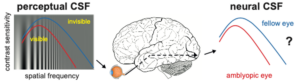
In a recent 7T MRI study at the Spinoza Centre for Neuroimaging, neuroscientists examined vision in people with amblyopia at both the behavioral and brain-level. Participants looked at simple visual patterns with one eye at a time, while the researchers measured brain activity using the 7 Tesla MRI scanner, with the goal of using the increased SNR at 7 Tesla to identify detailed differences in brain activation. In a separate visual task, they tested how well the people with amblyopia could see differences in contrast. In their results, the team found clear and consistent differences between the amblyopic eye and the unaffected eye, both in visual performance and in brain responses. These results show that the visual difficulties experienced by people with amblyopia are linked to changes in how the brain processes visual information.
This study was carried out by Carlien Roelofzen, Marcus Daghlian, Maartje de Jong, and Serge Dumoulin.